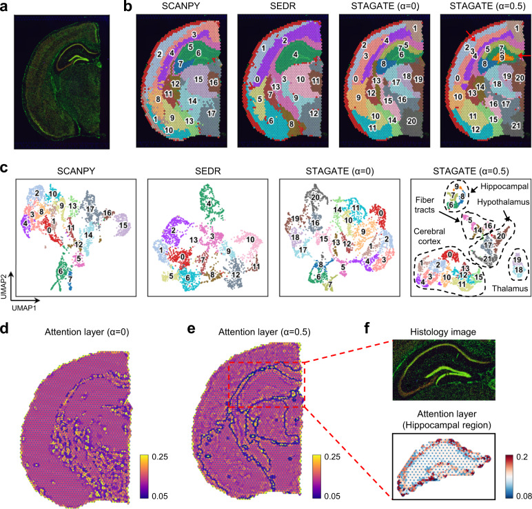
Fig. 5. STAGATE reveals spatial domains in adult mouse brain section profiled by 10x Visium.
a Immunofluorescent imaging of the tissue section stained with DAPI and Anti-NeuN. b Spatial domains generated by Louvain clustering with resolution = 1 on the low-dimensional embeddings of SCANPY, SEDR, STAGATE, and STAGATE with the cell type-aware module. The α represents the weight of the cell type-aware SNN (see Fig. 1). c UMAP visualizations of the low-dimensional embeddings of SCANPY, SEDR, STAGATE, and STAGATE with the cell type-aware module respectively. d, e Visualizations of the attention layer of STAGATE without (d) or with (e) the cell type-aware module. The nodes of the attention layer are arranged according to the spatial position of spots. The edges of the attention layer are colored by corresponding weights. f Zoomed-in views of immunofluorescent imaging of the hippocampus region and the visualization of attention layer in e.

