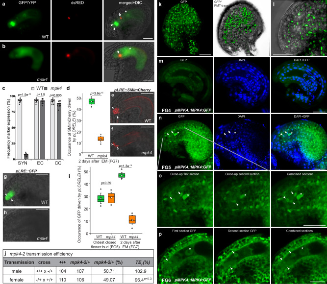
Fig. 2. Premature synergid degeneration at the molecular level, and MPK4 localization in the embryo sac.
a–c Expression of the female-gametophyte reporter (FGR7.0) in (a) WT and (b) mpk4. c Frequency of FGR7.0/+ expression in WT (n = 321) and mpk4 (n = 285) ovules was determined. Error bars show ± SD. d–i Expression of the LORELEI-reporter (pLRE) coupled to the Golgi-retention peptide sequence SMImCherry (d–f) and a single GFP (g–i). The expression of pLRE::GFP/- was analyzed in the oldest-closed flower bud (FG5 stage) (WT, n = 326; mpk4, n = 240) and 48 h after emasculation (WT, n = 328; mpk4, n = 251; FG7 stage). pLRE::SMImCherry/- was analyzed 48 h after emasculation (WT, n = 293; mpk4, n = 257). Boxes represent the 25th and 75th percentiles, and the inner rectangle highlights the median, whiskers show the SD, and outliers are depicted by dots (Min/max range). j Transmission analysis of mpk4. Transmission efficiency (TEf) indicates the transmission of mpk4 through the male and female gametophyte. k–p Representative images of MPK4:GFP expression and localization in the sporophytic ovule tissue (k–l) and at the female gametophytic developmental stages FG4, FG5, FG6 (m–p). k–l Depicted are maximum projections of Z-stacks, with a focal plane distance of 2 µm within a total range of about 30 µm, animated 3D-projection is provided in Supplementary movie 5. m–p pMPK4::MPK4:GFP-expressing line was analyzed, accompanied by the nuclei-staining dye DAPI. Negative control Supplementary Fig. 1l. Experiments were repeated three times with similar results and presented are representative images. c, d, i, j Statistical significance was analyzed by one-way ANOVA. Source data and further statistical analysis are provided in the source data file. White arrowhead, synergid; black arrowhead, degenerating synergid; white arrow, egg cell; white asterisk, central cell nucleus; black asterisks, polar nuclei. Scale bars, 20 µm (a, b, e, f–h), 30 µm (k–p).

