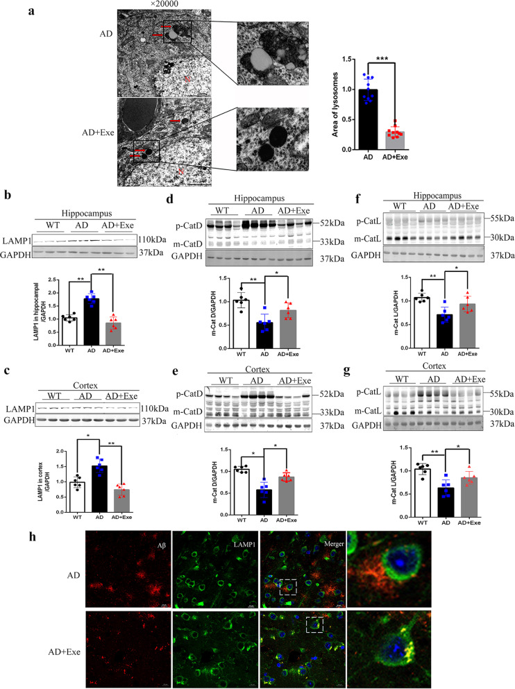
Fig. 4. Exercise activates lysosomal function and increases the colocalization of lysosomes with Aβ.
a Representative electron micrographs of lysosomes in pyramidal neurons from the CA1 area of the hippocampus from AD and AD + Exe mice. Arrows indicate lysosomes. (Scale bar, 1 μm). b, c Western blot of LAMP1 in hippocampus and cortex. d, e Maturation of cathepsin D in the hippocampus and cortex was assessed by Western blotting. f, g Maturation of cathepsin L in the cortex was assessed by Western blotting. GAPDH was used as a loading control (n = 4). h Representative colocalization of immunofluorescence image of Aβ and LAMP1. (Scale bar = 5 μm). Data represent means ± SD. *P < 0.05, **P < 0.01. One-way ANOVA test.

