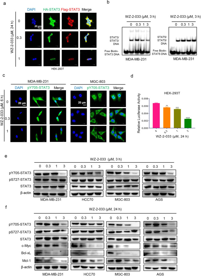
Fig. 2. WZ-2-033 disrupted STAT3 dimerization and inhibited STAT3 signaling.
a HEK-293T cells stably expressing HA-STAT3 (green) and Flag-STAT3 (red) proteins were treated with the indicated concentrations of WZ-2-033 for 24 h and assessed by immunofluorescence analysis. DAPI nuclear staining is shown in blue. Scale bar = 20 μm. b EMSA analysis: The nuclear extracts of MDA-MB-231 cells treated with WZ-2-033 were incubated with a biotin-STAT3 or biotin-STAT5 probe and subjected to EMSA. c Cellular localization of pY705-STAT3 (green) in the cells treated with WZ-2-033 for 6 h. DAPI nuclear staining is shown in blue. Scale bar = 20 μm. d STAT3C, pGL3-STAT3-promoter, and Renilla luciferase (as a reference) plasmids were transiently cotransfected into HEK-293T cells. The luciferase activity of the cells after treatment with WZ-2-033 for 24 h was detected as described in the methods. e The phosphorylation level (pY705 and pS727) and expression level of STAT3 in TNBC and gastric cancer cells were measured by Western blot after treatment with WZ-2-033 for 3 h. f The expression levels of c-Myc, Bcl-xL, and Mcl-1 in TNBC and gastric cancer cells were measured by Western blot after treatment with WZ-2-033 for 24 h.

