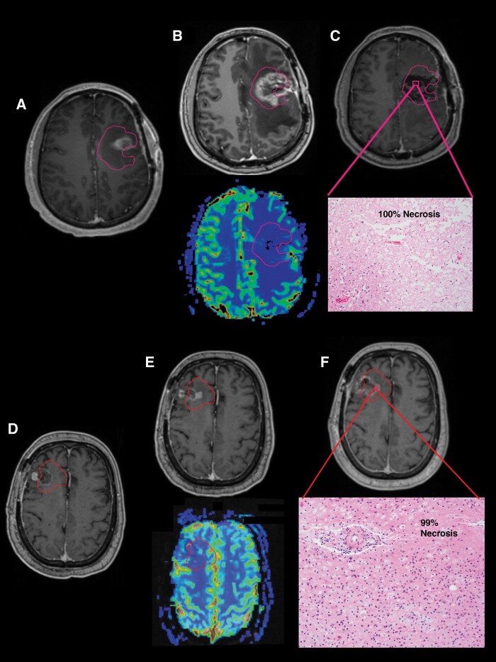
Figure 4.
Examples of 2 cases with “late pseudoprogression” or radiation necrosis are shown. Case 1 (A) At 1-month post-RT, residual enhancement is seen on T1w MRI within the pink PTV3 contour. (B) At 5-month post-RT, residual enhancement has increased, but the relative cerebral blood volume (rCBV) map on dynamic susceptibility contrast (DSC) perfusion MRI shows minimal to no perfusion. (C) Repeat resection demonstrated 100% necrosis on H&E-stained sections. Case 2 (D) At 1-month post-RT, there is primarily linear, postoperative enhancement on T1w-CE MRI within the red PTV3 contour. (E) At 8-month post-RT, increasing enhancement is seen at the periphery of the cavity. The DSC perfusion MRI again shows no definitive hyperperfusion. (F) Repeat resection again found predominantly (99%) necrosis on H&E-stained sections.

