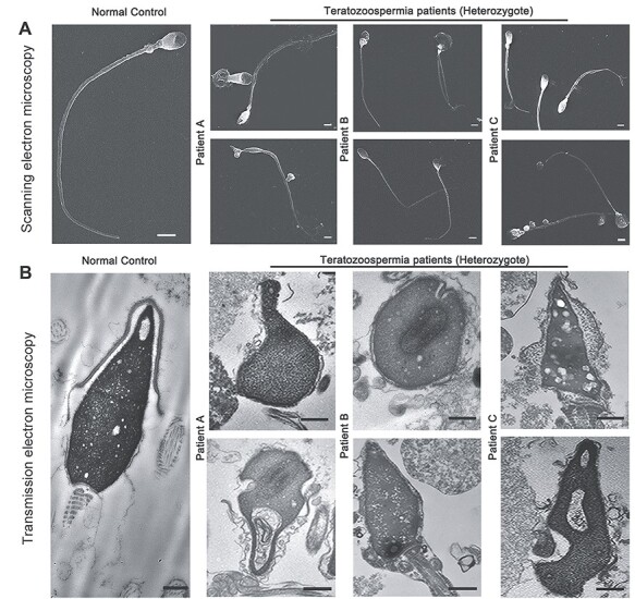
Figure 2.

The morphology of spermatozoa from the teratozoospermia patients by electron microscopy. (A) The abnormal sperm phenotypes were observed using SEM (scale bars, 5 μm). (B) TEM shows abnormal ultrastructure of the head from the patients’ spermatozoa compared to normal control (scale bars, 100 nm).
