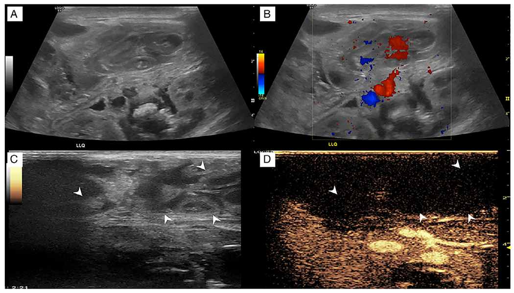
Fig. 10. Necrotizing enterocolitis.

A 1-day-old formerly premature girl was born at 29 weeks with gaseous distention on abdominal radiography. (a) Grayscale ultrasound in the left upper quadrant shows multiple dilated loops of bowel with wall thickening and hypoperistalsis to aperistalsis in real time. (b) Corresponding color Doppler image shows apparent flow in the mesentery but no appreciable flow in the bowel. However, the interpretation was limited by pulsatile motion from the patient’s high-frequency oscillator. (c, d) Dual-screen contrast-enhanced ultrasound displays loops of bowel (arrowheads) in the right upper quadrant that do not enhance (arrowheads) regardless of the high-frequency oscillator. (Images reprinted from Benjamin J et al. [31] with permission).
