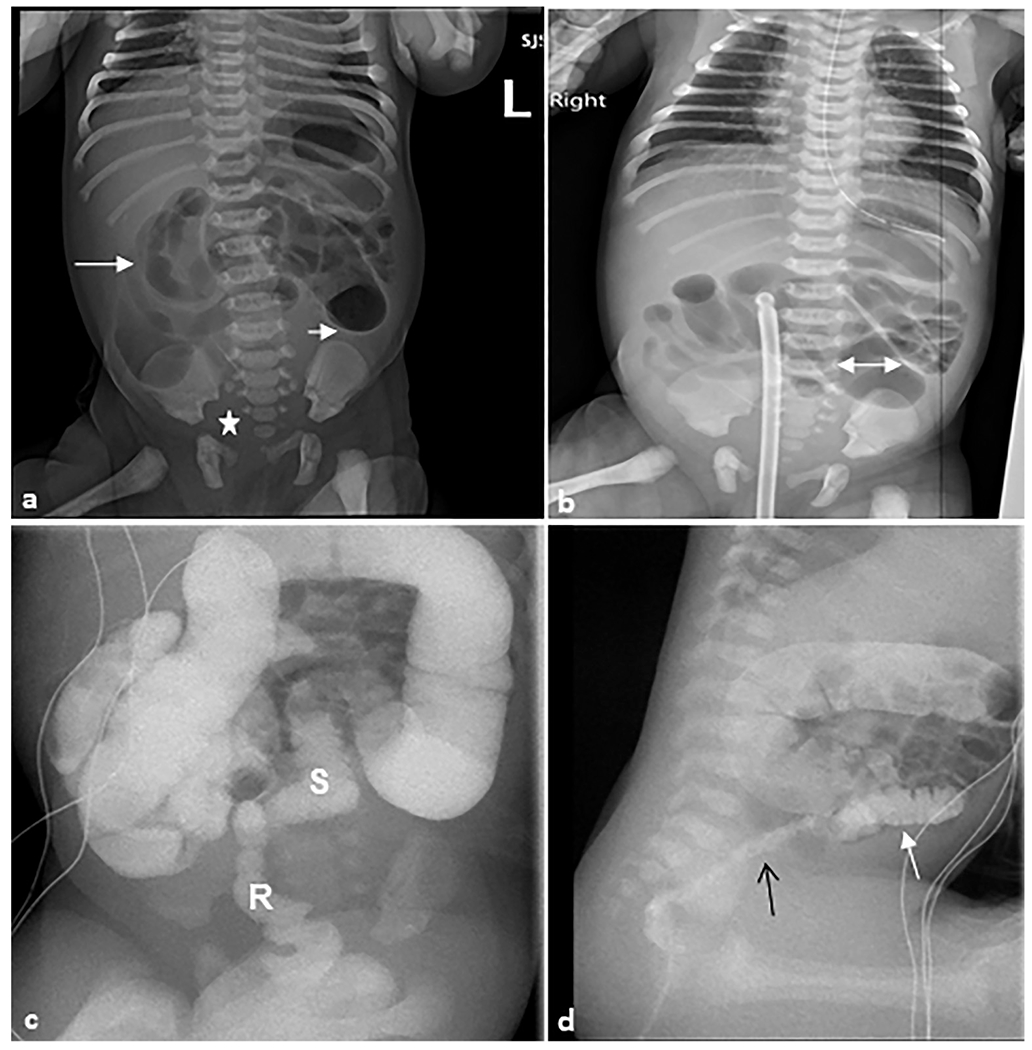
Fig. 13. Hirschsprung disease.

A 1-day-old female patient who presented with a distended abdomen and emesis. Rectal biopsy confirmed Hirschsprung disease. (a) Anteroposterior abdominal radiograph demonstrating multiple loops of air distended bowel (arrows) with paucity of bowel gas within the pelvis (star). (b) There are dilated proximal bowel loops with the maximal diameter of 2.7 cm (double-headed arrow) with featureless appearance of bowel loops in the right upper quadrant. (c) Fluoroscopic contrast enema demonstrating the sigmoid colon and rectum are small (S) in caliber up to the descending colon-sigmoid colon junction. A rectal catheter (R) is present with tip to the right of L2–L3. (d) Sagittal fluoroscopic image demonstrating an abnormal rectosigmoid ratio (<1) with the rectum (black arrow) of much smaller caliber than the sigmoid colon (white arrow) and upstream colon.
