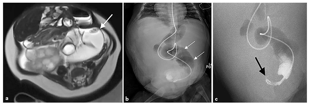
Fig. 5. Midgut volvulus.

A 4-month-old female patient with a history of Beckwith–Wiedemann syndrome presented with a distended abdomen. (a) Axial T2-weighted magnetic resonance image of the abdomen demonstrates a distended loop of bowel in the left abdomen (arrow) with an apparent twist in the mesentery. (b) Anteroposterior abdominal radiograph demonstrating multiple dilated loops of bowel with an enteric tube terminating in the pelvis. (c) Coronal upper GI series with contrast passed through the enteric feeding tube and revealed a “bird’s beak” sign (arrow), indicating twist around mesenteric axis. Subsequent exploratory laparotomy revealed jejunal volvulus.
