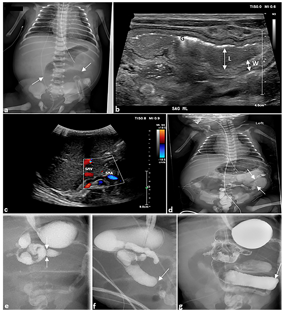
Fig. 6. Jejuno-ileal atresia.

Ex-premature infant. (a) Anteroposterior chest and abdomen radiograph (“babygram”) of a 1-day-old ex-premature infant born to parents with cystic fibrosis presenting with bilious emesis. The radiograph demonstrates dilated bowel loops (arrows). (b) Sagittal grayscale ultrasound image of the midline abdomen shows a dilated (L) loop of bowel with thickened walls (W). Echogenic foci are intraluminal gas (G). (c) Axial grayscale ultrasound of the right upper quadrant with color Doppler demonstrates the superior mesenteric vein and artery in normal position arguing against malrotation with midgut volvulus. (d) Water-soluble contrast enema demonstrates microcolon (arrows). (e) Upper gastrointestinal series demonstrates a normal duodeno-jejunal junction in the left upper quadrant (arrows) followed by abrupt cutoffs of the jejunum (arrows) in (f) sagittal and (g) anteroposterior views. At surgery, this was determined to be jejunal atresia, type IIIB.
