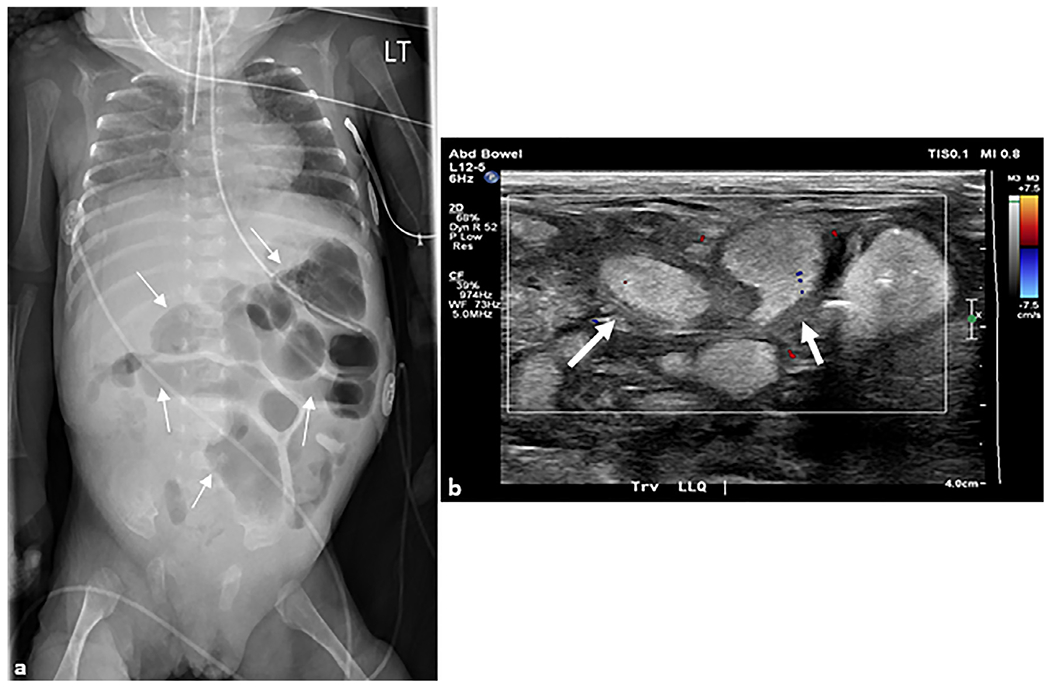
Fig. 8. Necrotizing enterocolitis.

(a) Anteroposterior chest and abdomen radiograph (“babygram”) of a 2-month-old ex-premature infant who presented to the intensive care unit with a tense and distended abdomen. Multiple distended loops of bowel (arrows) are seen in the left abdomen with a paucity of bowel in the right lower quadrant. (b) Transverse grayscale ultrasound image with color Doppler overlay of the right lower quadrant demonstrates multiple loops of distended small bowel containing complex fluid with thickened walls (arrows) and echogenic contents. No color Doppler flow is seen in the bowel wall concerning for ischemia. At surgery, this patient had 22 cm of ischemic bowel.
