Fig. 1.
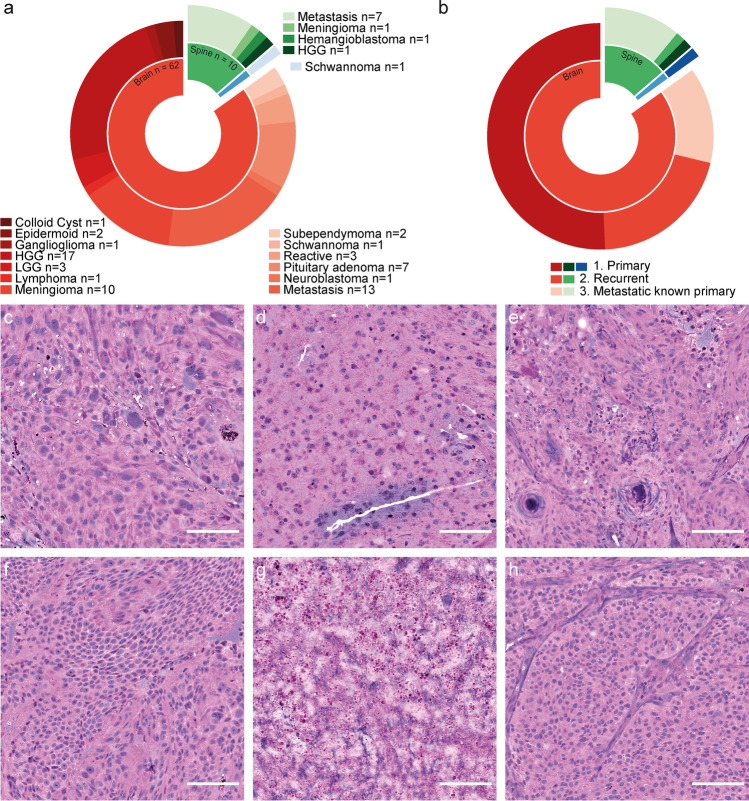
Data and stimulated Raman histology. a Category and location of 73 cranial, spinal, and peripheral (blue) tumors. b Distribution of patient history. c–h Illustrative examples of SRH images. c GBM of the left parietal lobe in a 72-year-old female. d 53-year-old male with left frontal oligodendroglioma WHO grade II. e Spinal (TH 2/3) psammomatous meningioma in a 78-year-old male. f Left frontal dural metastasis of an esophageal cancer in a 65-year-old male. g Reactive gliosis with necrotic components (shown) after radiation of a left temporo-occipital melanoma metastasis in a 40-year-old female. h Non-hormone active pituitary adenoma in a 56-year-old male. Scale bars, 100 µm. Program used to create figure: Adobe Illustrator CS 6

