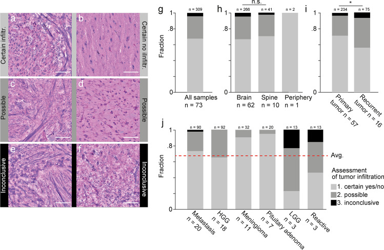
Fig. 2.
Assessment of tumor infiltration using SRH imaging. a–f Examples of subjective classification of tumor infiltration in SRH images as certain (a, b), possible (c, d), and inconclusive (e, f). a Certain tumor infiltration in case of a 78-year-old female with metastasis of NSCLC in the right frontal lobe. b Certain absence of tumor infiltration in case of cortical access tissue for resection of a right temporo-occipital GBM. c 72-year-old male patient with spinal metastasis of laryngeal squamous cell carcinoma. d 49-year-old male with recurrent left frontal GBM. e 77-year-old female with recurrent left temporal NSCLC metastasis. f 40-year-old female with metastasis of malignant melanoma in the left temporo-occipital lobe. g Overall assessment of tumor infiltration in 309 SRH images from 73 neurosurgical cases (cf. Fig. 1a). h Stratification of assessment of tumor infiltration according to tumor location and i the medical history. j Stratification of assessment of tumor infiltration according to diagnostic category (cf. Fig. 1a). Shown here are the 6 categories that contained > 3 cases and > 10 SRH images per category. Red line shows overall average (cf. Fig. 2g). Above all bars are the number of SRH images, below the number of cases per category. Scale bars, 100 µm. Program used to create figure, Adobe Illustrator CS 6

