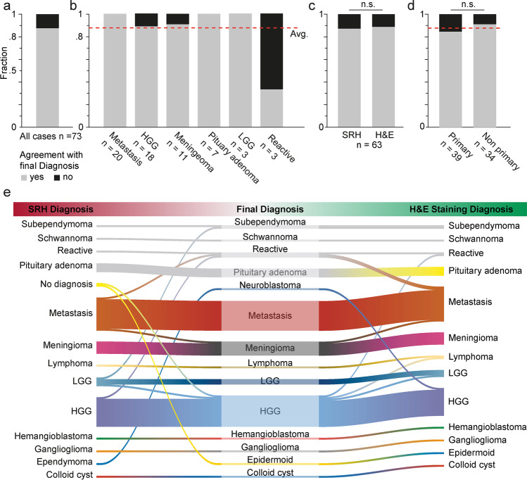
Fig. 3.
Accuracy of diagnosis based on medical history and SRH images. A board-certified neuropathologist novice in the assessment of SRH images stated a diagnosis based on the SRH images and the clinical information in 73 cases of cranial, spinal, or peripheral tumors. a Overall agreement of the diagnosis compared to the final neuropathological diagnosis was 87.7% (cf. red line in b and d). b Stratification of diagnostic accuracy according to tumor entity. Shown here are the 6 entities that contained > 3 cases. Below all bars are the number of patients per category. c Non-inferiority of diagnostic accuracy of SRH vs. conventional fast frozen section using H&E staining (87.3 vs. 88.9%, p = 0.783 chi-squared). d Non-significant lower accuracy in primary tumor cases (cf. group 1, Fig. 1b) vs. non-primary cases (cf. groups 2 and 3, Fig. 1b) (84.6 vs. 91.2%, respectively; p = 0.395 chi-squared). e River plot showing the correspondence of SRH-based diagnosis (left) and H&E-stained fast frozen sections (right) to the definitive neuropathological diagnosis (middle) with misclassifications appearing as lane changes. Program used to create figure, Adobe Illustrator CS 6

