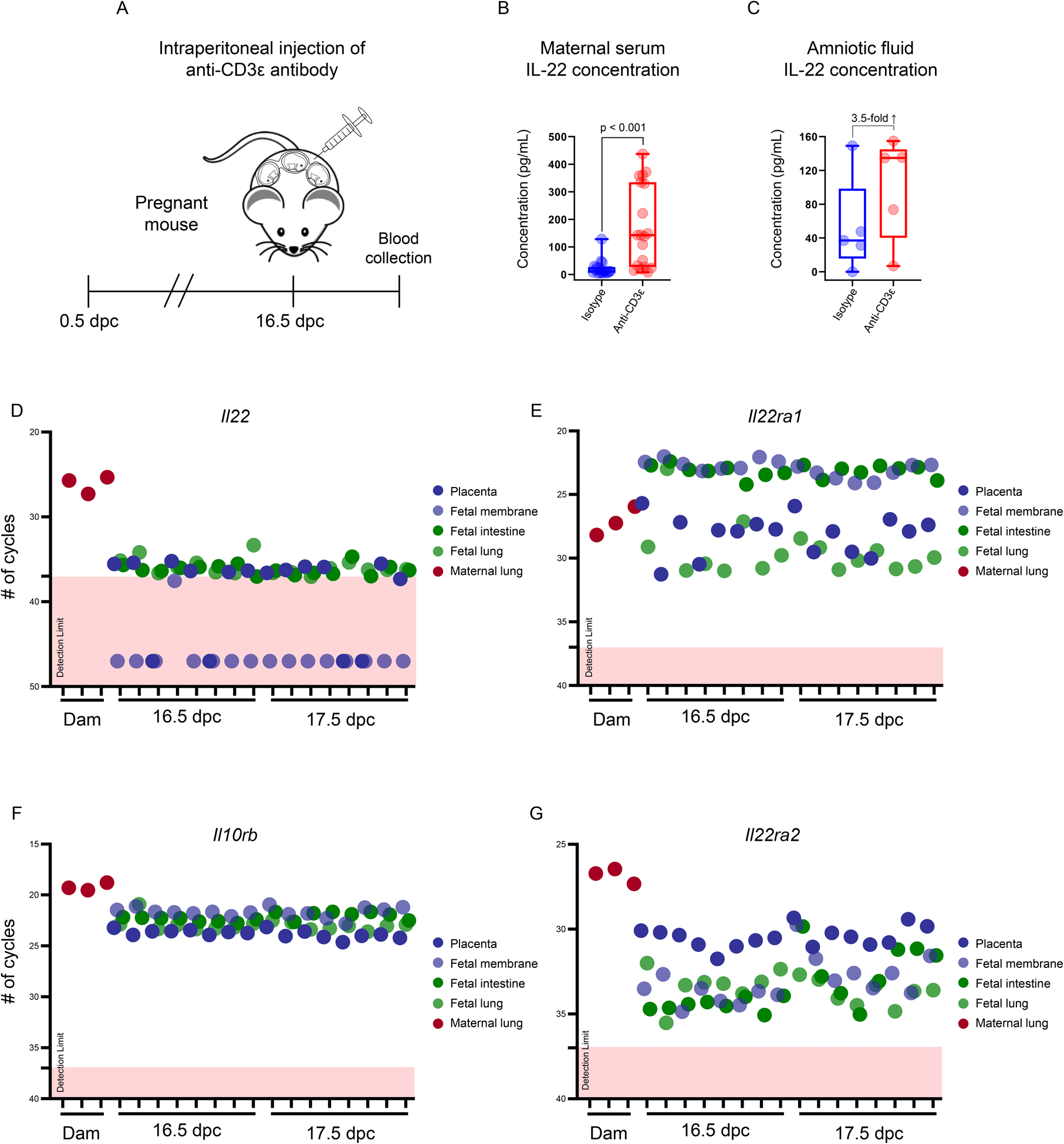
FIGURE 2. Maternal T-cell activation induces elevated IL-22 in the maternal circulation and amniotic cavity and the expression of IL22 and its receptors by gestational and fetal tissues in late gestation in mice.

(A) Study design illustrating the intraperitoneal injection of an anti-CD3ε antibody on 16.5 days post coitum (dpc) to induce T-cell activation-induced preterm birth in mice. Maternal serum and amniotic fluid were collected 12 – 16 h after injection for IL-22 determination. (B) Concentrations of IL-22 (pg/mL) in the maternal serum of mice injected with anti-CD3ε (n = 19) or isotype control (n = 18). (C) Concentrations of IL-22 (pg/mL) in the amniotic fluid of mice injected with anti-CD3ε (n = 5) or isotype control (n = 5). The p-values were determined using Mann-Whitney U-tests. Data are shown as scatter plots with medians, interquartile ranges, and min/max ranges. Dot plots representing the expression of (D) Il22, (E) Il22ra1, (F) Il10rb, and (G) Il22ra2 in the murine placenta, fetal membranes, fetal intestine, and fetal lung at 16.5 and 17.5 dpc, as well as the lungs of pregnant mice injected with lipopolysaccharide (positive control) at 17.5 dpc. Limits of detection for each transcript are denoted by dotted lines and pink boxes.
