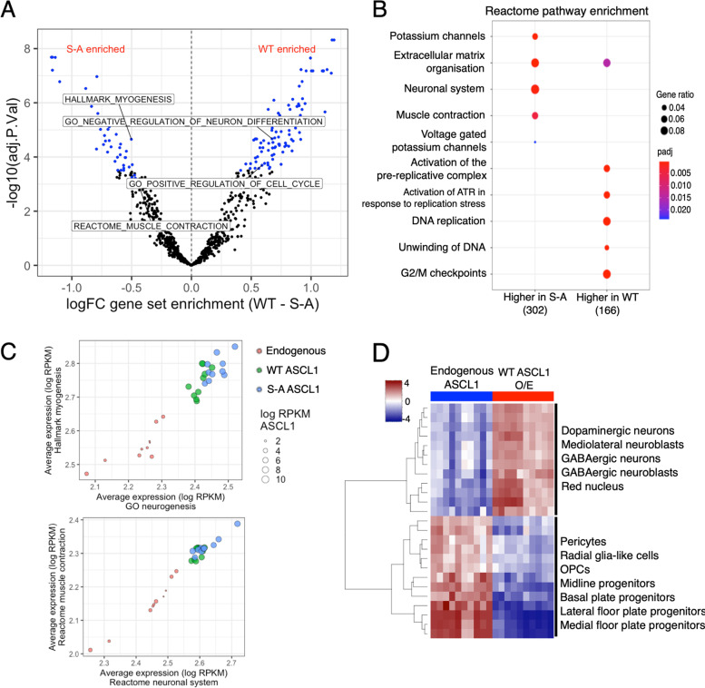
Fig. 4.
WT ASCL1 supports cell proliferation while un(der)phosphorylated ASCL1 promotes neurogenesis. a Volcano plot showing gene set variation analysis (GSVA), comparing RNA-seq data from WT and S-A ASCL1 overexpressing cells. Significantly differentially enriched terms highlighted in blue, terms of particular interest are labelled (padj < 0.05, n = 10) Gene sets preferentially enriched after WT or S-A ASCL1 induction have positive or negative enrichment values, respectively. b Dotplot showing significantly overrepresented Reactome Pathway gene sets. Gene sets were tested against genes significantly differentially expressed between WT and S-A ASCL1 overexpressing NB cells (DESeq2 padj < 0.05, n = 10). Total number of genes in each group shown in brackets. Point colour = Benjamini–Hochberg adjusted p-value, point size = gene ratio (number of genes related to term/total number of significant genes). c Dotplots showing the correlation of averaged expression of genes in the GO neurogenesis, and Hallmark myogenesis gene sets (left), or Reactome muscle contraction and neuronal system gene sets (right). Red = endogenous WT uninduced ASCL1, green = WT ASCL1, blue = S-A ASCL1. Point size indicates log2 RPKM ASCL1 expression, each point represents one biological replicate (n = 10). d Heatmap showing neuronal cell type marker gene sets (Broad Institute C8 set) found to be differentially enriched between WT uninduced and WT ASCL1 induced expression datasets (n = 10), by gene set variation analysis (GSVA)

