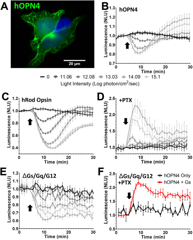
Fig. 1.
Human melanopsin shows light-dependent coupling of both Gαi/o and Gαs. (A) Immunocytochemistry photomicrograph showing human OPN4 (green) in HEK293T cells (DAPI-stained nuclei, blue). (B–F) Changes in bioluminescence in response to a 1 s 470 nm light flash (arrow), normalised to 1 at the time of the light pulse, from HEK293 cells expressing the cAMP reporter GloSensor and either hOPN4 (B,D–F) or hRod Opsin (C). (B–E) Responses across a range of flash intensities for hOPN4 (B) and hRod Opsin (C) in HEK293T cells, hOPN4 in HEK293T cells treated with pertussis toxin (PTX) to eliminate Gi/o signalling (D) and hOPN4 in HEK293 ΔGs/Gq/G12 cells (E). Key for light intensity in B–E is shown below B (B and C, n=4; D and E, n=3). (F) hOPN4-expressing HEK293 ΔGs/Gq/G12 cells treated with PTX with (hOPN4+Gs) or without (hOPN4 only) heterologous Gαs exposed to a 1s 14.09 log photon/cm2/sec light flash (hOPN4 only, n=3; hOPN4+Gs, n=5). Data expressed as mean±s.e.m. Cells pretreated with 2 µM forskolin to elevate the starting level of cAMP for traces in B,C,E. NLU, normalised luminescence units. n values denote biological replicates from independent transfections.

