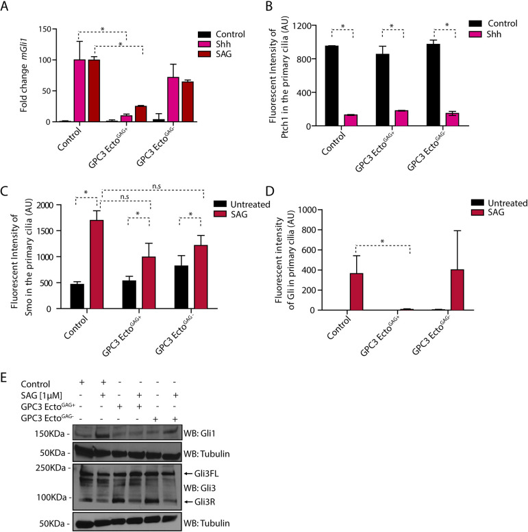
Fig. 3.
Purified GPC3 ectodomain inhibits Hh signaling as dominant negative. (A) Wild-type MEFs treated with GPC3-EctoGAG+ or GPC3-EctoGAG− were incubated with SAG (1 µM) or Shh for 24 h, and Hh pathway output was measured by qRT-PCR for Gli1. GPC3-EctoGAG+ inhibits Hh signaling in wild-type MEF cells. Data are mean±s.e.m. of three replicates. (B) Wild-type MEFs stably expressing Ptch1-eGFP were incubated with Shh for 24 h in the presence or absence of 1 µM of purified GPC3-EctoGAG+ or GPC3-EctoGAG−, and ciliary intensity of Ptch1 was measured by immunofluorescence microscopy. Cilia were detected by staining for endogenous Arl13B. GPC3-EctoGAG+ and GPC3-EctoGAG− had no effect on Shh-induced Ptch1 exit from cilia. Data are mean±s.d. (300-400 cilia were measured per condition). (C) As in B but treating wild-type MEFs with SAG (1 µM) and measuring the intensity of endogenous Smo in primary cilia. GPC3-EctoGAG+ and GPC3-EctoGAG− had no effect on SAG-induced ciliary accumulation of Smo. Data are mean±s.d. (300-400 cilia were measured per condition). (D) As in C but measuring the intensity of endogenous Gli proteins in primary cilia. GPC3-EctoGAG+ abolishes Gli recruitment to ciliary tips by Hh pathway activation, whereas GPC3-EctoGAG− has no effect. Data are mean±s.d. (300-400 cilia were measured per condition). (E) As in D but cells were analyzed by immunoblotting for Gli1 and Gli3. Blotting for tubulin served as a loading control. GPC3-EctoGAG+ did not block the reduction in Gli3R levels caused by Hh pathway activation but blocks the accumulation of Gli1. Blots shown are representative of three experiments. *P<0.05; n.s., not significant (two-tailed paired t-test). AU, arbitrary units.

