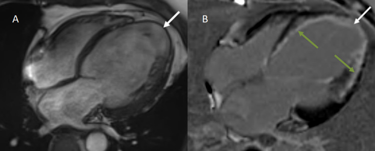
Figure 6. A: Four-chamber CINE TRUFI sequence depicting mild cardiomegaly with dilated left atrium and LV. Severe thinning of the cardiac apex (1 mm) with ballooning is also noted (white arrow). B: Delayed post-contrast enhanced imaging in the four-chamber view in the same patient demonstrates transmural (>50%) LGE in the apex (white arrow) and subendocardial (<50%) LGE involving the interventricular septum and lateral wall of the LV (green arrows).
LGE, late gadolinium enhancement; LV, left ventricle

