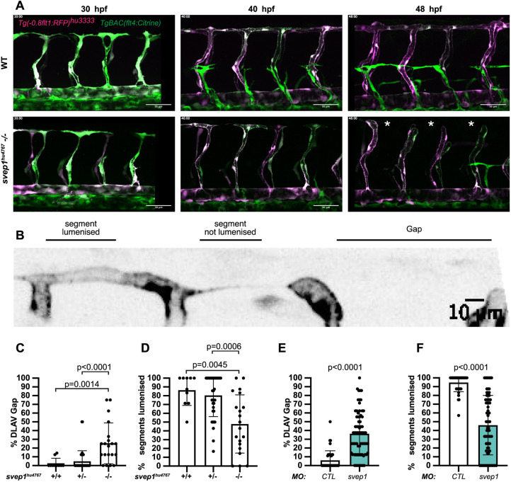
Fig. 1.
svep1 mutant and morphant zebrafish embryos exhibit vascular anastomosis defects. (A) Stills from time-lapse movie of Tg(-0.8flt1:RFP)hu3333; TgBAC(flt4:Citrine) embryos injected with MO-CTL (5 ng) or MO-svep1 (5 ng) and treated with 1× (0.014%) tricaine from 30 to 48 hpf. White asterisks indicate gaps in the DLAV. (B) Still from a time-lapse movie of a Tg(-0.8flt1:RFP)hu3333; TgBAC(flt4:Citrine) embryo injected with MO-CTL (5 ng) exhibiting a gap in the DLAV between two adjacent ISVs. Side view, dorsal side left. (C) Bilateral quantifications of the percentage of gaps in the DLAV at 48 hpf in WT (n=5), svep1hu4767 heterozygous (n=18) and svep1hu4767 homozygous embryos (n=10) treated with 1× tricaine (0.014%) from 30 to 48 hpf (N=3). (D) Bilateral quantifications of the percentage of lumenised segments in the DLAV at 48 hpf in WT (n=5), svep1hu4767 heterozygous (n=18) and svep1hu4767 homozygous embryos (n=10) treated with 1× tricaine (0.014%) from 30 to 48 hpf (N=3). (E) Bilateral quantifications of the percentage of gaps in the DLAV at 48 hpf in embryos injected with MO-CTL (5 ng) (n=20) and MO-svep1 (5 ng) (n=36) and treated with 1× tricaine (0.014%) from 30 to 48 hpf (N=6). (F) Bilateral quantifications of the percentage of lumenised segments in the DLAV at 48 hpf in embryos injected with MO-CTL (5 ng) (n=20) and MO-svep1 (5 ng) (n=36) and treated with 1× tricaine (0.014%) from 30 to 48 hpf (N=6). Data are mean±s.d. Mann–Whitney test. Scale bars: 50 μm (A); 10 μm (B).

