Fig. 6.
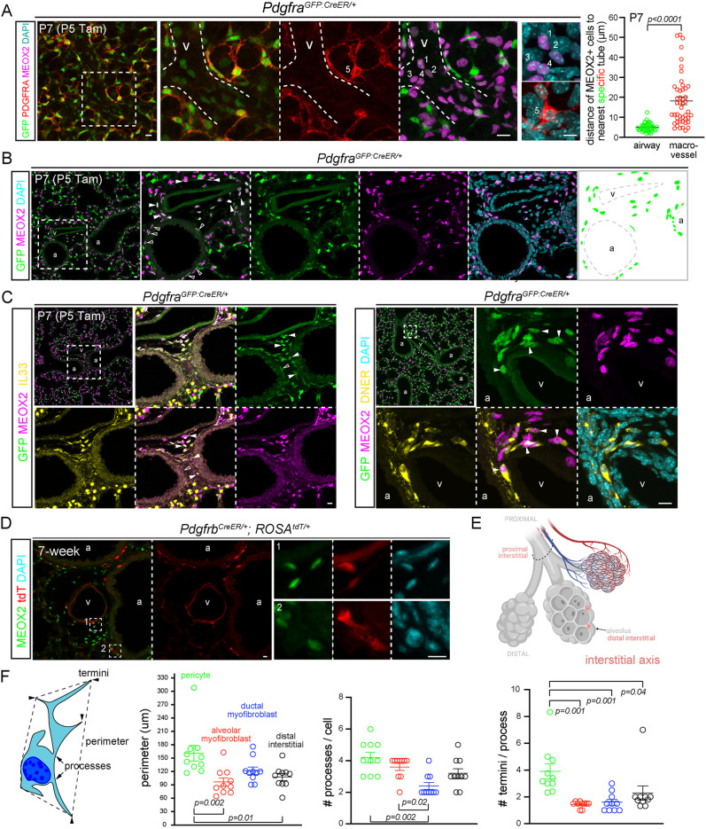
Proximal interstitial cells are in bronchovascular bundles and express MEOX2/IL33/DNER. (A) Left: dim GFP, MEOX2+ cells (1-4) around a vessel (curved dashed lines) and bright GFP, PDGFRA-high cells (5). Dashed box in the first image indicates area shown at higher magnification in the following images in all instances. Tamoxifen (Tam) facilitates nuclear accumulation and, hence, detection of the GFP:CreER fusion protein. Right: nearest neighbor analysis of perpendicular distance of MEOX2+ cells to the basement membrane of the respective tubes. The large spread of the vessel category is from MEOX2+ cells in vascular adventitia (unpaired Student's t-test; data are mean±s.d.). See Fig. 6B,C for representative images. (B) Immunostaining images and diagram showing GFP+, MEOX2+ cells around airways (open arrowhead) and vessels (filled arrowhead) within the bronchovascular bundle. (C) GFP+, MEOX2+ cells (filled arrowhead) are IL33+ (left) and DNER+ (right) within bronchovascular bundles. Occasional GFP+ cells are MEOX2− (open arrowhead). (D) Besides VSM cells, PdgfrbCreER labels MEOX2+ cells within bronchovascular bundles. Tamoxifen 3 mg was administrated 2 days before lung harvest. (E) Proximal interstitial cells within the bronchovascular bundle (dashed semioval). Created with BioRender.com. (F) Schematic and quantification of cell morphology of color-coded cell types in Fig. 2C (P3 pericytes), Fig. 3D (P7 alveolar myofibroblasts), Fig. 4E (P21 ductal myofibroblasts) and Fig. 5E (6-week distal interstitial cells) (ordinary ANOVA with Tukey test; data are mean±s.d.). Each symbol represents a cell; the number of termini per process is averaged over all processes of a given cell. Scale bars: 10 µm. a, airway; Tam, 300 µg Tamoxifen; v, vessel.

