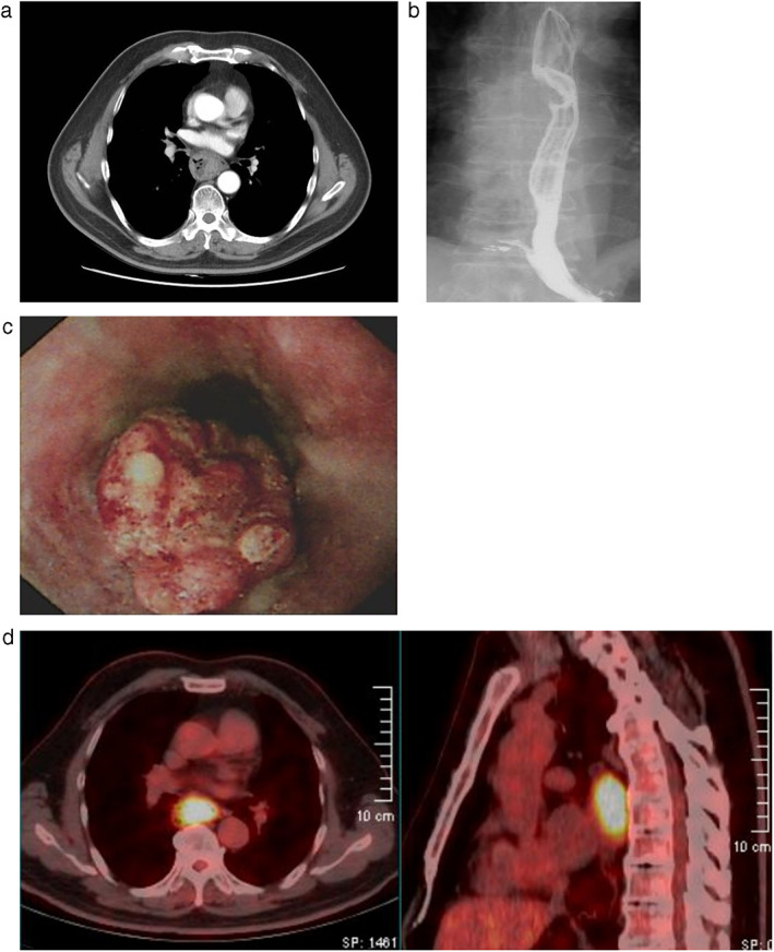
FIGURE 1.

(a) Contrast‐enhanced chest computed tomography demonstrates thickening of the middle esophageal wall. (b) Barium swallow shows a filling defect of the middle esophagus. (c) Endoscopy reveals a 4.0 cm mid‐esophageal mass. (d) PET/CT image shows a mid‐esophageal soft tissue mass with high FDG uptake without lymph node involvement
