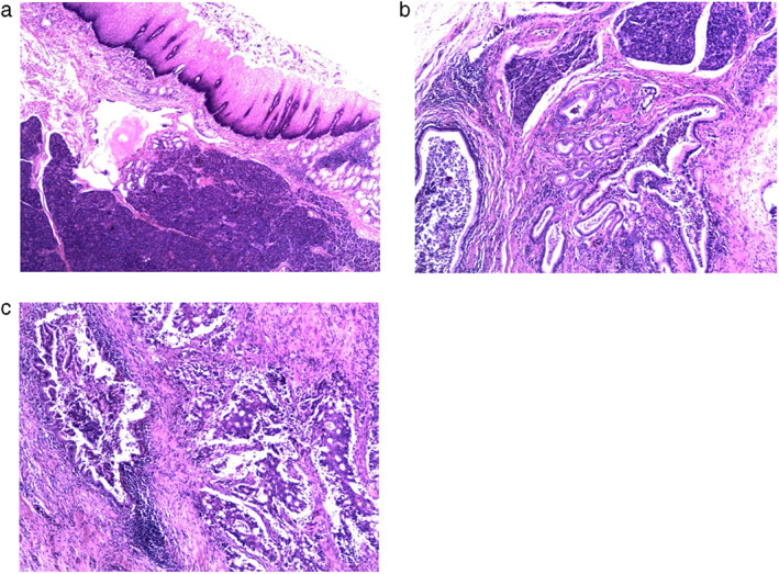
FIGURE 2.

(a) Ectopic pancreas with lobular structure highlighted in the submucosa and mucosa muscularis of the esophagus (×40 magnification). (b) Ectopic pancreas with lobular acini and hyperplastic ductal glands (×100 magnification). (c) Right‐sided moderately differentiated adenocarcinoma with cribriform structure. On the left side, a ductal gland was identified with pancreatic intraepithelial neoplasia 1 (PanIN‐1) changes next to the carcinomatous glands, which indicates that this adenocarcinoma originated from the pancreatic duct (×100 magnification)
