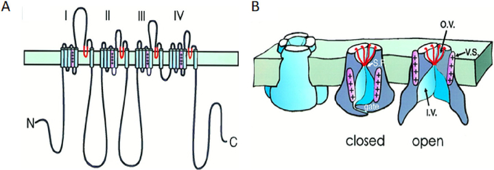
Fig. 2.
Cartoons from the 1990s of a four-fold pseudosymmetric voltage-gated cation channel in the plasma membrane. Peptide chain folding (A) and functional components (B) are shown diagrammatically with selectivity filter regions in red and the voltage sensor S4 segment in pink. The drawing in B was made before structural work showed where the voltage sensors lie. They are drawn too close to the pore itself and should be shifted more laterally virtually into the lipid membrane (see Fig. 4). The folding diagram is for an Na+ channel with four homologous repeat domains labeled I-IV. Ca2+ channels would be similar, and K+ channels are homo- or hetero-tetramers of subunits similar to a single Na+ channel domain. Abbreviations: S.F., selectivity filter; v.s., voltage sensor; o.v., outer vestibule; and i.v., inner vestibule. (A) After Catterall (1992) and (B) from Armstrong and Hille (1998).

