Figure 1.

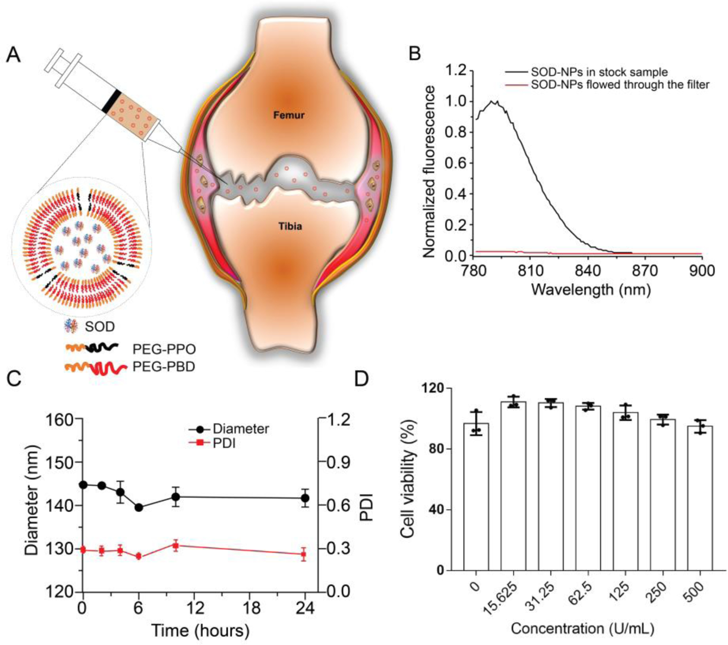
Preparation and characterization of SOD-NPs. (A) Schematic diagram of SOD-loaded polymersomes with high membrane permeability for intra-articular joint injection. (B) Evaluation of SOD retention within PEG-PPO-doped polymersomes in PBS buffer (0.1 M, pH 7.4). The liquid that flowed through the filter was measured for fluorescence (red line). The fluorescence of unfiltered sample in the presence of Triton X-100 was also recorded (black line). The fluorescence intensity is normalized relative to the intensity of unfiltered sample at 790 nm. (C) The stability of SOD-NPs in bovine synovial fluid was accessed by monitoring the hydrodynamic diameter and PDI for up to 24 hours. (D) The cytotoxicity of SOD-NPs was determined by measuring the cell viability of primary chondrocytes after coincubation with SOD-NPs at various SOD concentrations.
