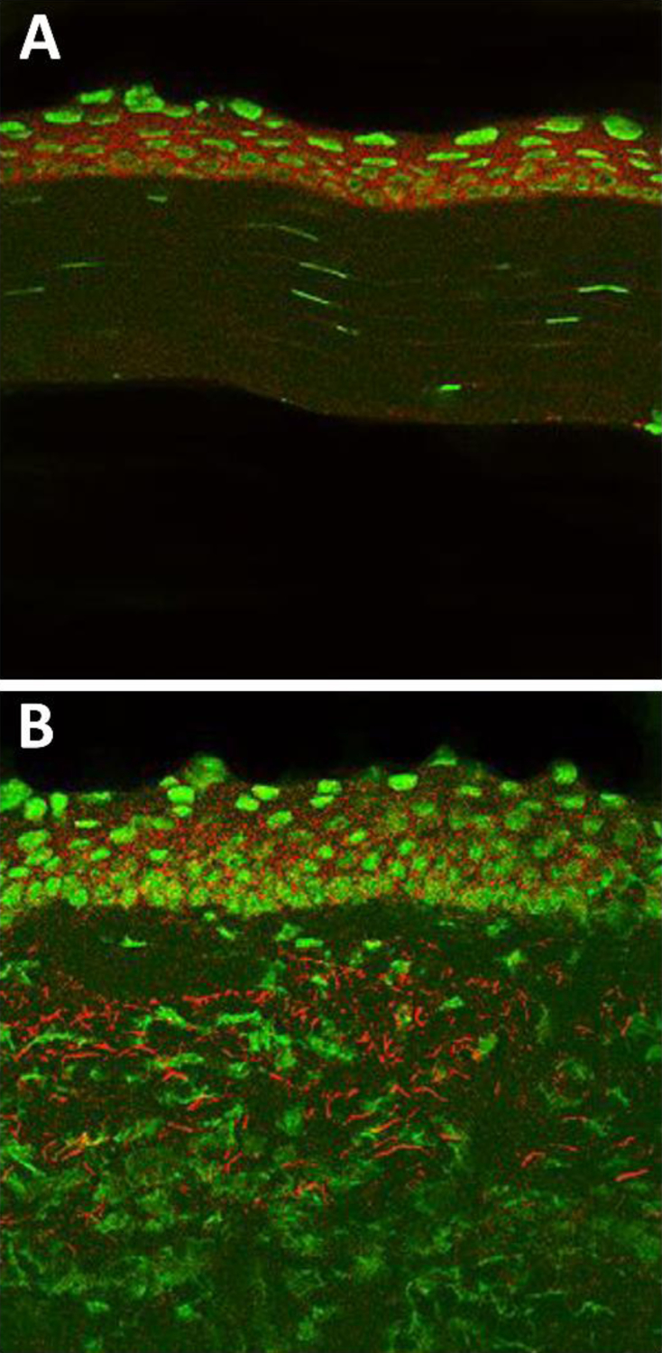
Figure 2.

Normal and 5 day post infection C57BL/6 mouse cornea stained with an anti-HMGB1 Ab. (A) HMGB1 epithelial staining (red) in normal, uninfected cornea. (B) HMGB1 staining distributed throughout the epithelium and stroma after 5 days of infection. Nuclear marker=Sytox Green.
