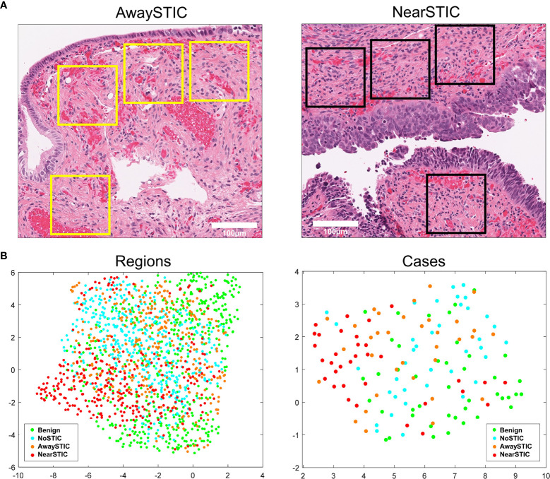
Figure 1.
The fallopian tube stromal architecture changes both locally and globally in the presence of STIC lesions. (A) Yellow boxes illustrate stromal regions where tiles were extracted away from the STIC lesion (left panel) and black boxes illustrate stromal regions where tiles were extracted near the STIC lesion (right panel). (B) Low-dimensional UMAP representation of stromal morphometric features in regions and cases with benign fallopian tubes (Benign), uninvolved fallopian tubes from patients with HGSOC (NoSTIC), and fallopian tubes with STIC lesions and HGSOC. Image regions from fallopian tubes with STIC lesions were stratified based on proximity to the lesion (awaySTIC and nearSTIC). Expression in an individual case represents median expressions from regions in that case. The UMAP plots were generated using the run_umap.m function in MATLAB.

