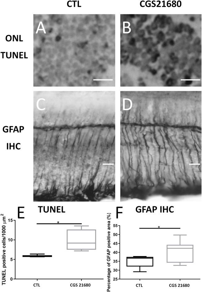
FIGURE 1.
Treatment with CGS21680 increased number of apoptotic nuclei and GFAP immunoreactive areas. (A,B) Representative sections showing TUNEL staining of ONL of retina of a CTL eye (A) and a CGS21680-treated eye (B). Scale bar: 20 μm. (C,D): GFAP-immunostained sections from retina of a CTL eye (C) and a CGS21680-treated eye (D). More intense GFAP immunoreactivity of Müller cells is observed in retina of CGS21680-treated eye compared with CTL. (E) Quantification of ONL TUNEL-positive cells. CGS21680 produced a significant increase in positive nuclei of ONL compared with CTL (Student’s t-test, *p < 0.05). (F). Quantification of GFAP immunoreactive area. Boxes represent 25 and 75 percentiles, whiskers represent minimum and maximum values, and transverse lines represent medians. CGS21680 produced a significant increase in expression of GFAP compared with CTL (Student’s t-test, *p < 0.05).

