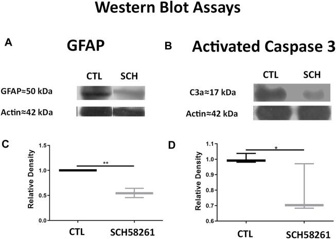
FIGURE 6.
Treatment with SCH58261 decreased levels of GFAP and activated caspase-3. (A) Representative Western blot (WB) for GFAP of CTL and SCH58261-treated eyes. (B) Representative Western blot for activated caspase-3 of CTL and SCH58261-treated eyes. (C) Quantifications of GFAP WB of CTL and SCH58261-treated eyes (D). Quantification of activated caspase-3 WB of CTL and SCH58261-treated eyes. Relative densities were normalized against CTL β-actin. Boxes represent 25 and 75 percentiles, whiskers represent minimum and maximum values, and transverse lines represent medians. Student’s t-test, *p < 0.05; **p < 0.01.

