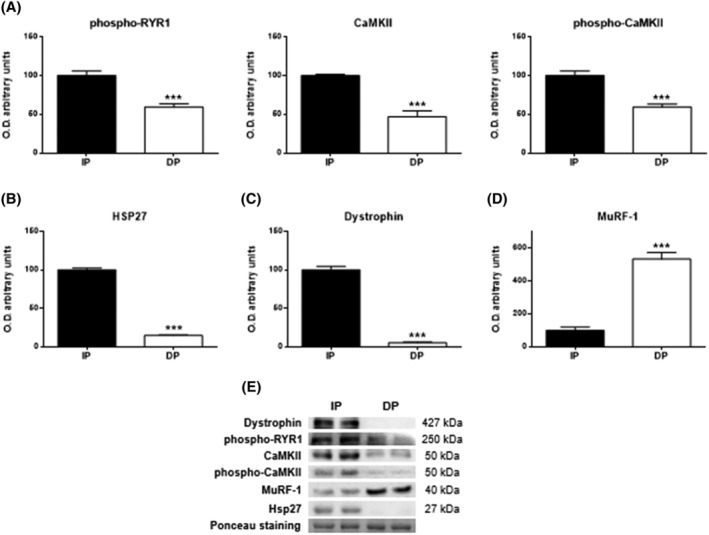
Figure 5.

Skeletal muscle excitation‐contraction coupling mechanism in muscle tissues of functional‐independent patients (IP) and functional‐dependent patients (DP). (A) Levels of phospho‐RYR1, Ca2+/calmodulin‐dependent protein kinase II (CaMKII) and phospho‐CaMKII. (B) Levels of Hsp27, (C) Dystrophin, and (D) MuRF‐1. Bar chart shows the quantification of the optical densities (O.D.). (E) Representative immunoblots. Ponceau staining was used as a loading control. Data are represented as the mean ± SEM. ***P < 0.001.
