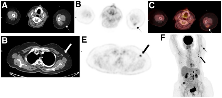
FIGURE 2.
A 52-y-old woman with gastrointestinal tumor was referred for routine 18F-FDG PET/CT follow-up study. Study was performed 2 d after second COVID-19 vaccination. Selected transaxial CT (A and D) and PET (B and E) slices at level of posterior arm uptake and axillary LNs, fused image (C) at level of posterior arm uptake, and maximum-intensity projection (F) demonstrate moderate-intensity uptake in left posterior arm (thin arrows) (SUVmax, 3.6) and high-grade activity in left axillary nodes measuring 1.0 cm in short axis with benign appearance (thick arrows) (SUVmax, 7.1). Maximum-intensity projection also shows high-grade 18F-FDG activity in retroperitoneal nodes and multiple implants.

