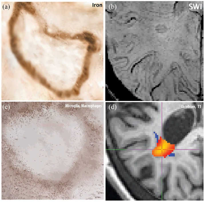
Figure 1.
Chronic active lesion pathology-imaging features: Panel (a) shows a cartoon of the iron deposition at the edge of a chronic active lesion and panel (b) shows an example of a hypointense rim on a susceptibility-weighted scan probably reflecting iron. Panel (c) shows a cartoon of activated microglia/macrophages in the periphery of a chronic active lesion and we assume that this inflammatory activity is responsible for low expansion of SEL lesion visible in (d).

