Figure 1.

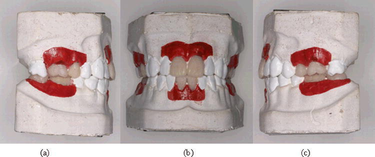
Dental arch model with resin teeth of anatomical root form placed at the mesial and distal of the six surgery sites. Red area represents the attached gingiva. (a) Right posterior, (b) anterior, and (c) left posterior areas.

Dental arch model with resin teeth of anatomical root form placed at the mesial and distal of the six surgery sites. Red area represents the attached gingiva. (a) Right posterior, (b) anterior, and (c) left posterior areas.