
FIGURE 2.
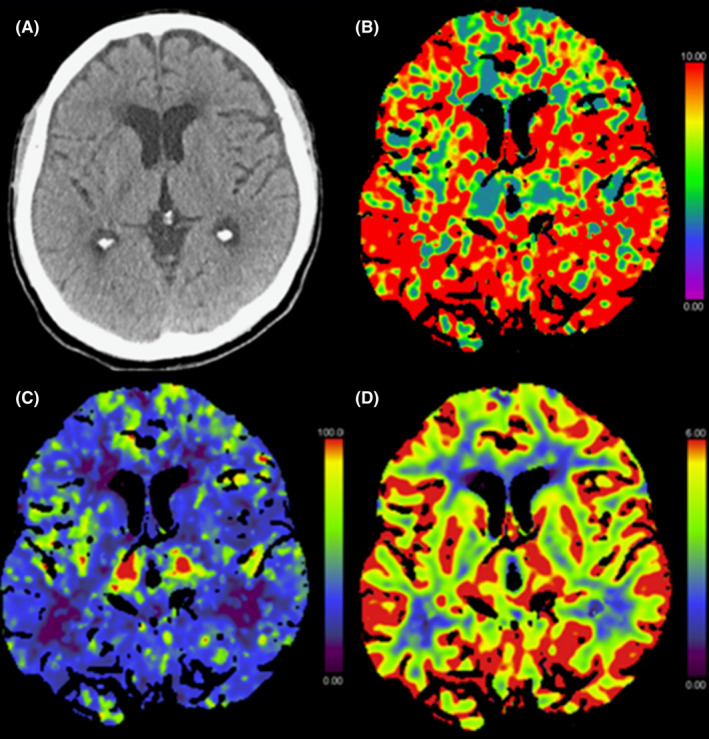
Initial axial non‐enhanced computed tomography (CT) (A) showing normal parenchyma with no evidence of swelling or ischemic changes. CT perfusion showing prolonged mean transit time (MTT) (B) with global cortical hypoperfusion including the basal ganglia in the cerebral blood flow map (C), but no abnormalities in the cerebral blood volume map (D)