Fig. 1.

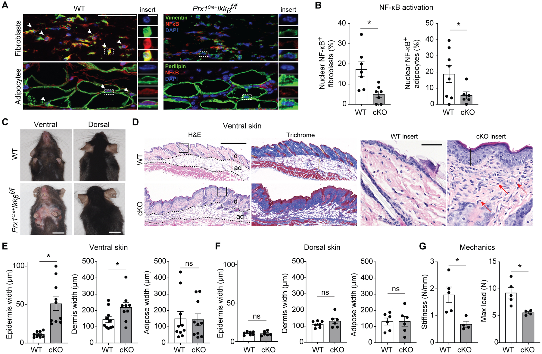
Preventing basal NF-kB activation in Prx1+ mesenchymal cells leads to ventral skin anomalies. A. Representative immunofluorescent images of ventral skin stained with NF-kB (p65) antibody and vimentin (fibroblast marker) or perilipin (adipocyte marker) in 16-week-old control (WT) or experimental (Prx1Cre+Ikkbf/f) mice. White arrows point to fibroblasts or adipocytes with nuclear NF-kB expression. Scale bar, 50um; insert scale bar, 5um). B. Quantification of double-positive vimentin+/nuclear NF-kB+ fibroblasts per total vimentin+ cells (left), and perilipin+/nuclear NF-kB+ adipocytes per total perilipin+ cells (right), comparing between WT and Prx1Cre+Ikkbf/f (conditional knockout, cKO) group. n=7–8 mice in each group. C. Representative photograph images of ventral and dorsal skin in 16–20-week-old WT and Prx1Cre+Ikkbf/f mice; scale bar, 1cm. D. Hematoxylin and eosin (H&E) and trichrome images of ventral skin from WT or cKO mice; d, dermis, ad, adipose layer. Scale bar, 0.5mm. Insert, red arrows point to eosinophils. Scale bar, 50um. E and F. Epidermis, dermis and adipose width in ventral (E) or dorsal (F) skin of WT and cKO mice. n=6–10 mice in each group. H. Mechanical testing of ventral skins from WT and cKO mice; stiffness and maximum load until failure (tear) were quantified. n=4–5 each. Data are represented as mean ± SEM of biological replicates. Animal experiments were repeated two to three times. *P<0.05; Student’s t-test comparing WT to cKO groups.
