
Fig. 4.
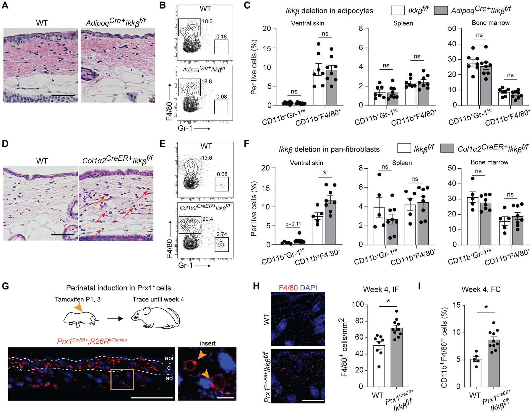
Ikkb deletion in skin fibroblasts, not adipocytes, is responsible for myeloid inflammation during perinatal growth. A. H&E images of ventral skin in 4-week-old control group (WT) or adipocyte-specific Ikkb deletion (Adipoq-Cre+Ikkbf/f) mice. Scale bar, 50um. B. Flow cytometry analysis of neutrophils (CD45+CD11b+Gr-1Hi) and F4/80+ myeloid cells (CD45+CD11b+F4/80+) from enzymatically digested ventral skin of WT or Adipoq-Cre+Ikkbf/f mice. C. Quantification of neutrophils and F4/80+ myeloid cells per total live cell count in ventral skin (left), spleen (middle), and bone marrow (right) comparing WT versus adipocyte-specific deletion of Ikkb group. n=7–8 each. D. H&E images of ventral skin in 4-week-old WT or experimental mice that had induced deletion of Ikkb in Col1a2-expressing fibroblasts (Col1a2CreERT+Ikkbf/f). Mice received tamoxifen twice at P1 and P3 (50ug/dose) and were euthanized at 4 weeks of age. Red arrows point at monocytic and/or eosinophilic cells. Scale bar, 50um. E. Flow cytometry plot for neutrophils and F4/80+ myeloid cells in WT or mice with Ikkb-deleted in Col1a2+ fibroblasts. Tamoxifen was administered at P1 and P3 (intragastric, 50ug/dose). F. Quantification of neutrophils and F4/80+ myeloid cells per total live cell count in ventral (left), spleen (middle) and bone marrow (right) comparing WT versus Ikkb deletion in Col1a2-expressing pan-fibroblasts. n=5–8 each. G. Perinatal induction of dTomato reporter gene in postnatal Prx1+ cells (Prx1CreERT+R26RdTomato), labeling dermal mesenchymal cells and some subcutaneous adipocytes (insert, arrows; scale bar, 20um) after 4-week tracing period. Scale bar, 0.5mm. H. Immunofluorescent images of F4/80+ cells in the ventral skin of 4-week-old WT mice and Prx1CreERT+Ikkbf/f mice that had perinatal deletion of Ikkb (left), and quantification of F4/80+ cells per mm2 (right). IF=immunofluorescence analysis. n=8–10 each. Both WT and Prx1CreERT+Ikkbf/f mice received tamoxifen. Scale bar, 50um. I. Flow cytometry analysis (FC) for F4/80+ myeloid cells from ventral skin of WT and Prx1CreERT+Ikkbf/f mice. n=5–10 each. Data are represented as mean ± SEM of biological replicates. Animal experiments were repeated three times. *P<0.05, ns=not significant; Student’s t-test comparing WT to each respective experimental group.