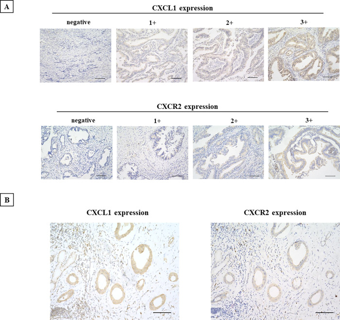
Fig 2. Representative immunostaining images of CXCL1 and CXCR2 expression.
A, CXCL1 was expressed in CCA cells. CXCR2 was observed at both the cell membrane and the cytoplasm. Intensity score: 0, negative; 1+, weakly positive; 2+, positive; 3+. Bar: 100 μm. B, CXCL1 and CXCR2 expression in a case. Bar: 100 μm.

