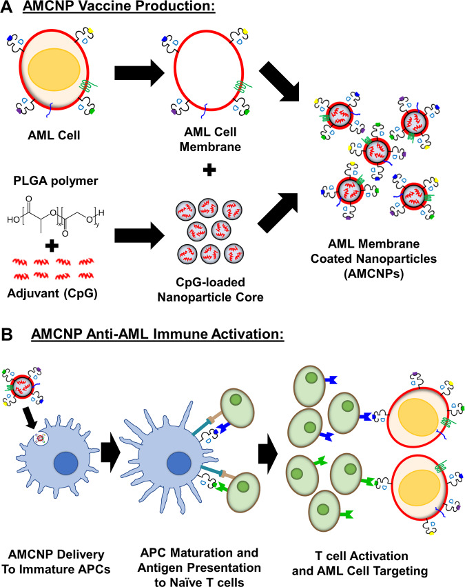
Fig. 1. Schematic of AML membrane-coated nanoparticles (AMCNPs) production and anti-leukemic vaccination.
A The immunostimulatory adjuvant, CpG oligodeoxynucleotide 1826, was encapsulated into biodegradable poly (lactic-co-glycolic acid) (PLGA) polymer nanoparticle cores (small gray spheres) via a double emulsion process. Through sonication, CpG-loaded nanoparticle cores were coated with isolated AML cell membrane (red circle), including membrane-associated MHC-I-presented antigens (blue, green, purple, and yellow dots), to form AMCNPs. B Delivery of AMCNPs to immature APCs (blue cell) stimulates maturation and AML-associated antigen presentation. The mature APCs (blue cell) present AML antigens and co-stimulatory molecules to naive T cells (green cells), resulting in activation and proliferation of T cells specific for different AML antigens (blue, green). Activated T cells (green cells) can initiate AML cell death, after detecting the MHC-I-presented antigens on AML cells (red cells).

