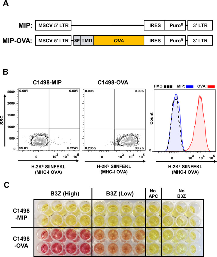
Fig. 2. Characterization of C1498-OVA cell line.
A MIP vector and MIP-OVA retrovirus constructs used in generation of C1498-MIP and C1498-OVA cell lines. The murine Cadm1 signal peptide (SP) and transmembrane domain (TMD) were cloned 5’ to full-length chicken ovalbumin into the MIP vector. B Representative flow cytometry plots and histogram of C1498-MIP and C1498-OVA cell lines stained with antibodies against MHC class I-presented OVA peptide 257–264 (H-2Kb:SIINFEKL), which demonstrate OVA antigen presentation in C1498-OVA cells compared to C1498-MIP or fluorescence minus one (FMO) negative control staining. C OVA-specific CD8+ T cell (B3Z) lacZ activation assay. C1498-MIP or C1498-OVA cells were incubated with B3Z CD8+ T cell hybridoma reporter cells, in which OVA-specific T cell receptor activation drives lacZ expression. Representative image demonstrating OVA-specific T cell activation (red color) in B3Z lysates, as assayed with the β-gal substrate chlorophenol red-β-galactoside (CPRG).

