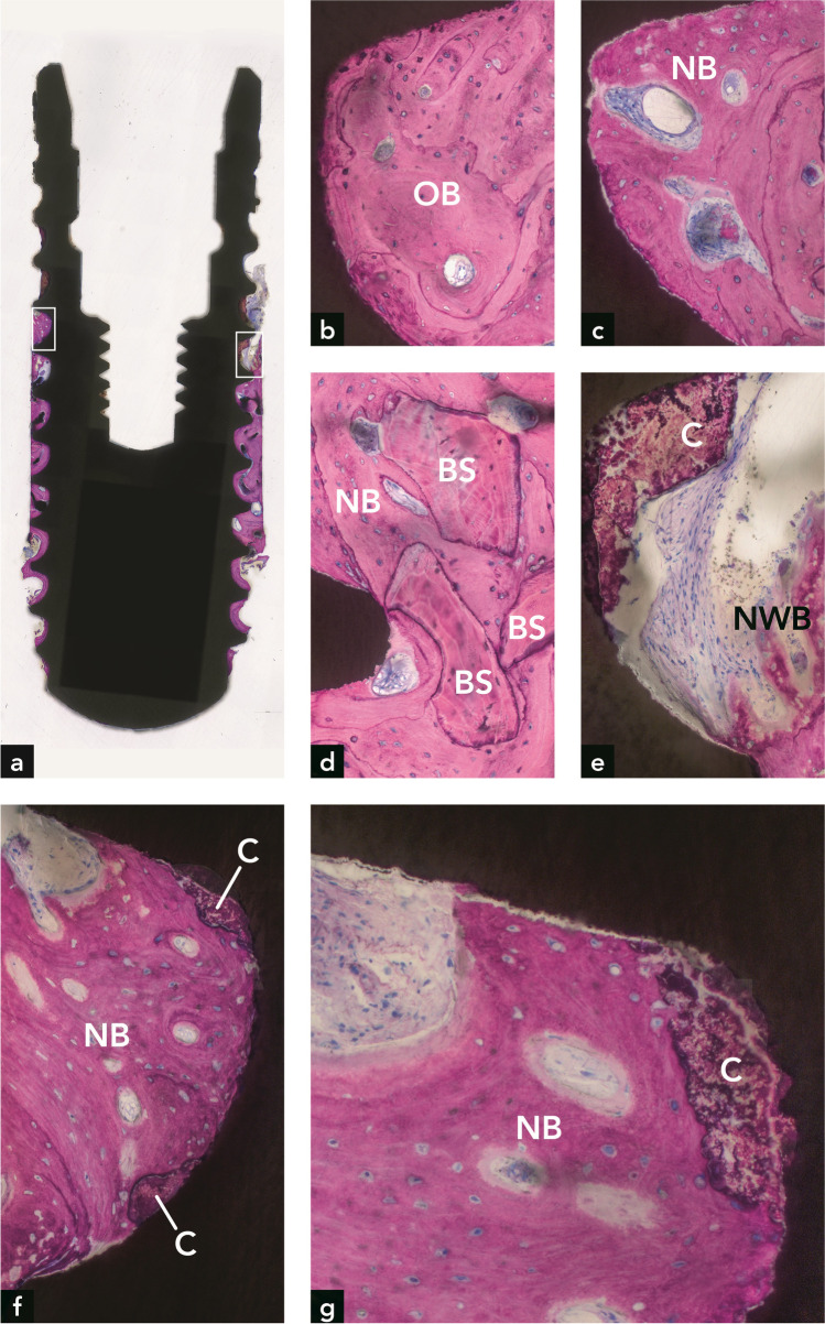
Fig. 3.
Implant #2. (a) Plenty of bone is seen around the implant. (b) Old bone (OB) is present in the apical part of the implant. (c) New bone (NB) is present in the coronal part of the implant. (d) Three bone substitute (BS) particles are embedded in new bone. (e) Higher magnification of the right rectangle in (a), illustrating new woven bone (NWB) and calculus (C) at the coronal termination of bone on the implant surface. (f) Higher magnification of the left rectangle in (a), illustrating new bone in direct contact with two calculus deposits on the implant surface. (g) Higher magnification of direct contact between new bone and calculus in an adjacent section

