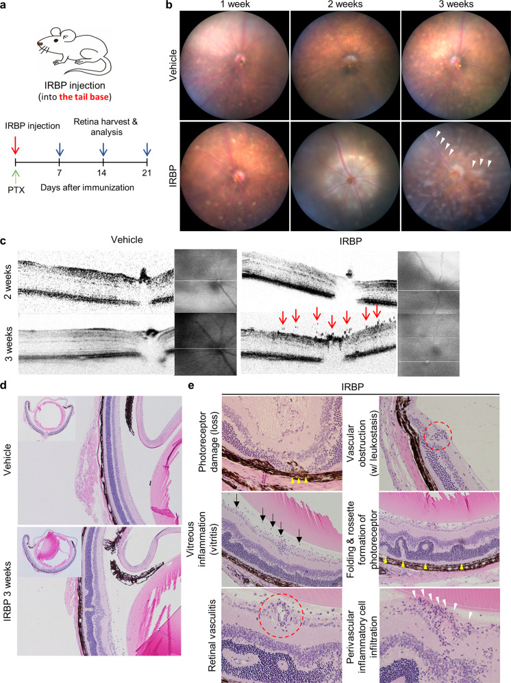
Fig. 1. Interphotoreceptor retinoid-binding protein (IRBP)-immunized mice present perivascular inflammation.
a Schematic diagram of the experimental schedule used to contract the IRBP-induced experimental autoimmune uveitis (EAU) model. IRBP peptide emulsified in complete Freund’s adjuvant (CFA) was injected into the tail base, and adjuvant (purified Bordetella pertussis toxin) was administered intraperitoneally. b Fundus images of mice with EAU showing diffuse perivascular inflammation evidenced by vascular sheathing (white arrowhead). c. Optical coherence tomography (OCT) images of mice with EAU showing inflammatory cells on the preretinal surface and in the superficial retinal layers (red arrows). d, e Histologic features indicating retinal vasculitis in IRBP-immunized mice. d Hematoxylin and eosin staining of the eyecup and retinal layer in the EAU mouse model. e Representative images showing various signs of vascular inflammation and subsequent retinal damage in mice with EAU. The arrow, arrowheads, and red-dotted circles indicate areas representative of the different phenotypes.

