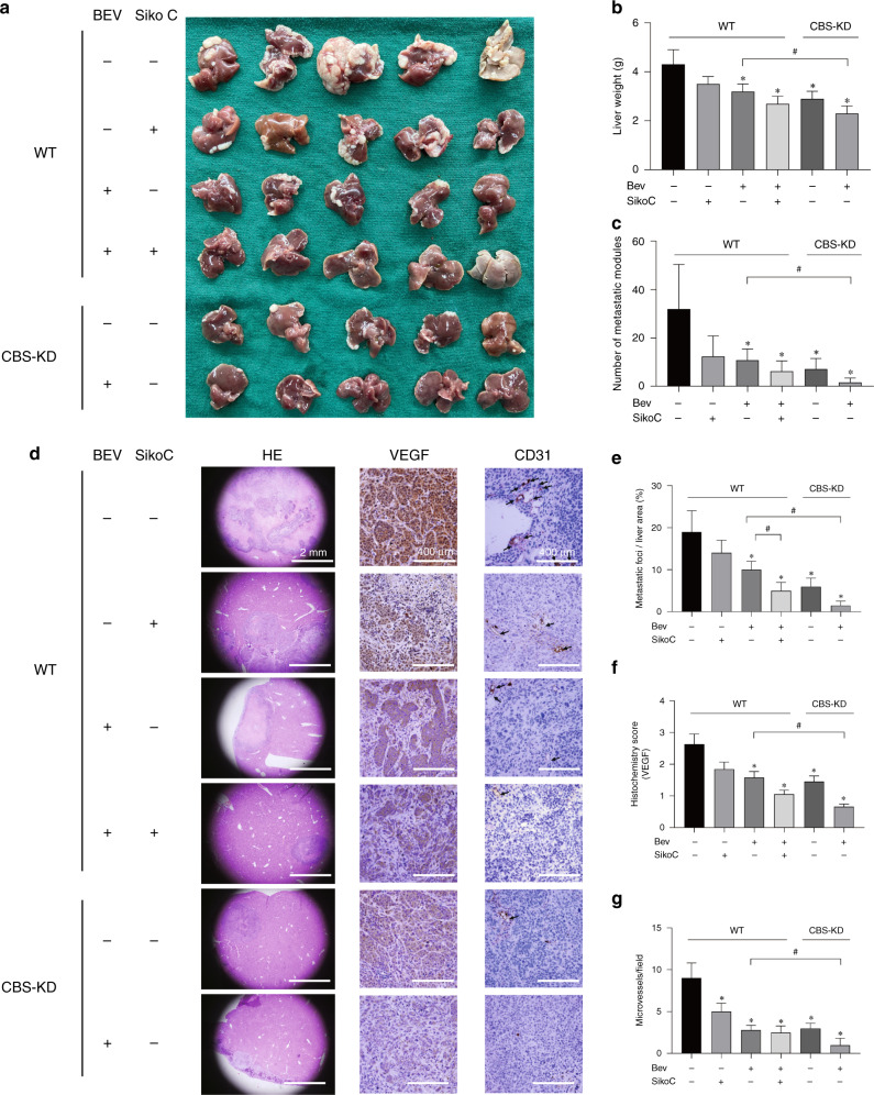
Fig. 5. The effect of CBS and VEGF on liver metastasis of colon cancer cells in vivo.
a Images of liver metastatic nodules 6 weeks after tumour cell injection. Bev represents bevacizumab. b Average mouse liver weights in the 6 groups. (n = 5). c Average number of metastatic nodules. d Representative images of HE and immunohistochemical staining in liver tissue sections from the 6 groups. e Average area of metastatic foci in liver sections from the 6 groups. f VEGF histochemical score in liver tissue sections from the 6 groups. g Average number of microvessels stained with CD31 in liver tissue sections from the 6 groups. *P < 0.05 vs. the control group. #P < 0.05 vs. the group of mice implanted with WT cells and treated with Bev (+) but not sikokianin C (−).

