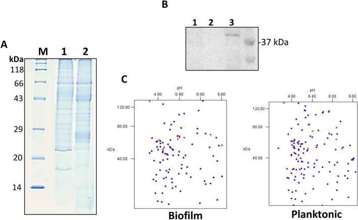
Figure 1.
Analysis of the proteome of P. intermedia biofilm and planktonic cells. (A) SDS–PAGE gel showing protein bands from protein preparations: biofilm (lane 1) and planktonic cells (lane 2). (B) Western blot analysis of the secretome preparations (lane 1 = biofilm, lane 2 = planktonic) and the WCP (lane 3) using an antibody for the cytoplasmic marker protein FtsZ. (C) Protein sequences from LC–MS analysis of the secretome were analyzed by an in silico 2DE tool.

