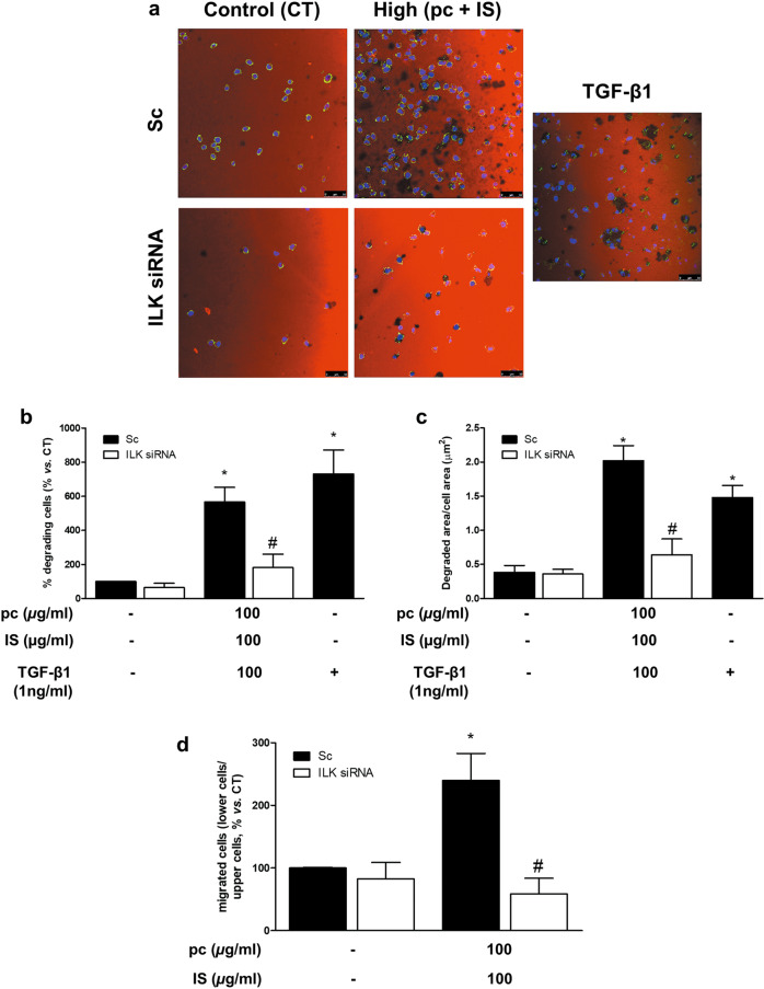
Fig. 4. p-Cresol (pc) and indoxyl sulfate (IS) induce THP-1 cell matrix degradation and cell migration.
THP-1 cells were transfected with scrambled RNA (Sc) as a control (CT) (upper microphotographs, b–d black bars) or were depleted of ILK with specific siRNA (lower microphotographs, b–d white bars). a–c Afterward, the cells were seeded on TRITC-gelatin-coated coverslips and incubated with high concentrations of pc plus IS for 24 h. a Confocal micrographs showing the distribution of TRITC gelatin (red) and THP-1 cells stained with phalloidin (green) and Hoechst 33342 (blue). The results of a representative experiment are shown. Scale bar, 50 µm. b, c Bar graphs indicating the average percentage of THP-1 cells with an associated subjacent area of gelatin degradation (b) or the total degraded area divided by the total cell area (µm2) (c) per field of view for cells treated as in a. d Afterward, the cells were loaded in the upper chamber of the filter and incubated with high concentrations of pc plus IS for 24 h. Cell migration was determined by Transwell migration assay. The bar graphs indicate the average percentage of THP-1 cells that migrated across the filter toward MCP-1 cells treated as in a. b, c The results are expressed as a percentage of the number of untreated CT cells. All values are presented as the mean ± SEM from 4 or 6 independent experiments. *P < 0.05 vs. CT; #P < 0.05 vs. Sc. TGF-β1 was used as a positive control. MCP-1 was used as a chemoattractant.

