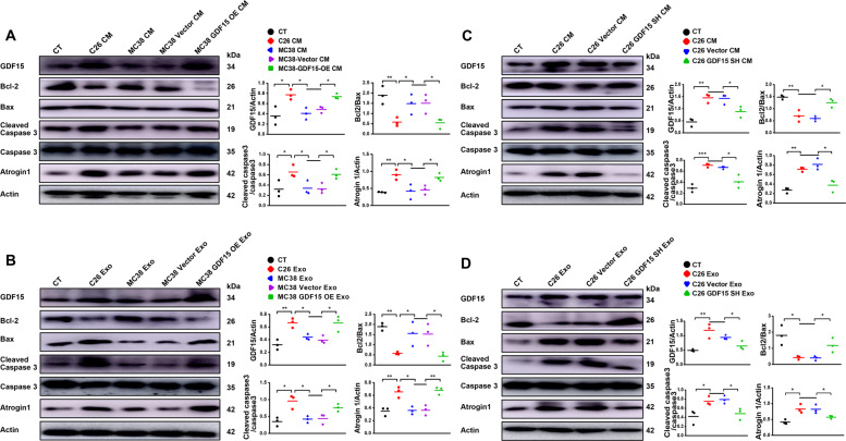
Fig. 7. Confirmation of the involvement of apoptotic pathway in GDF15-induced muscle atrophy.
C2C12 myotubes were treated with MC38-GDF15-OE cell medium, exosomes or C26-GDF15-SH cell medium, exosomes respectively. A Analysis of the protein level of MHC, GDF15, Bcl-2, Bax, Cleaved caspase-3, Atrogin1 in C2C12 myotubes treated with MC38-GDF15-OE cell medium (n = 3, *p < 0.05, **p < 0.01; t test). B Analysis of the protein level of MHC, GDF15, Bcl-2, Bax, Cleaved caspase-3, Atrogin1 in C2C12 myotubes treated with MC38-GDF15-OE cells derived exosomes (n = 3, *p < 0.05, **p < 0.01; t test). C Analysis of the protein level of MHC, GDF15, Bcl-2, Bax, Cleaved caspase-3, Atrogin1 in C2C12 myotubes treated with C26-GDF15-SH cell medium (n = 3, *p < 0.05, **p < 0.01, ***p < 0.001; t test). D Analysis of the protein level of MHC, GDF15, Bcl-2, Bax, Cleaved caspase-3, Atrogin1 in C2C12 myotubes treated with C26-GDF15-SH cells derived exosomes (n = 3, *p < 0.05, **p < 0.01; t test). Data presented are the mean ± SEM.

