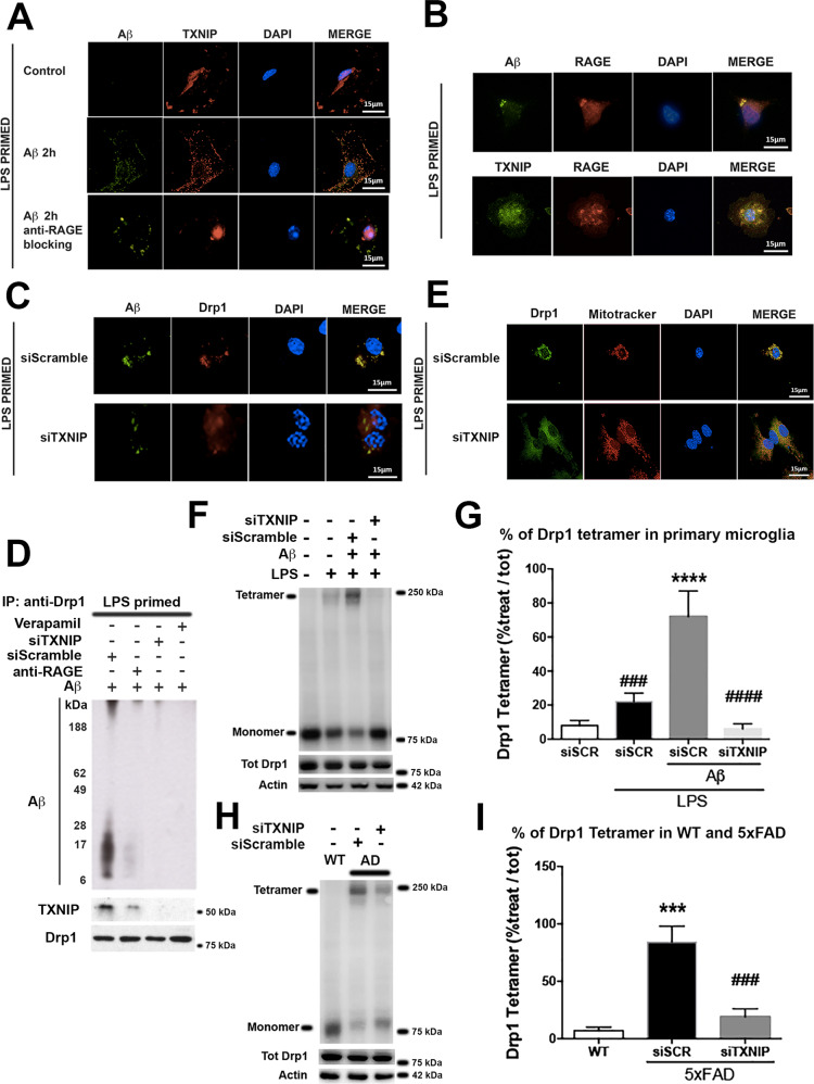
Fig. 3. TXNIP is required for Drp1- Aβ interaction and Drp1 oligomerization in LPS-primed primary microglia and 5XFAD Tg mice.
A IF analysis of Aβ (green) and TXNIP (red), nuclei are in blue (representative of n = 5). B IF analysis of Aβ (green), TXNIP (green), and RAGE (red), nuclei are in blue (representative of n = 5). C IF analysis of Aβ (green) and Drp1 (red), nuclei are in blue (representative of n = 3). D Western blot analysis of Aβ oligomers, TXNIP, and Drp1 after co-immunoprecipitation with an anti-Drp1 antibody (representative of n = 4). E IF analysis of Drp1 (green) and Mitochondria (red), in microglia, treated as indicated nuclei are in blue (representative of n = 3). F, G Western blot of native gel (F) and quantification (G) of Drp1 tetramer in LPS-primed primary microglia. Total Drp1 and actin were used to normalize. One-way ANOVA followed by Tukey’s multiple comparison test (n = 3; ****p < 0.0001 versus control; ###p < 0.001, ####p < 0.0001 versus siScramble). H, I Western blot of native gel (H) and quantification (I) of DRP1 tetramer in wt and 5xFAD mice treated as indicated. Total Drp1 and actin were used to normalize. One-way ANOVA followed by Tukey’s multiple comparison test (n = 3; ***p < 0.001 versus control; ###p < 0.001 versus siScramble).

