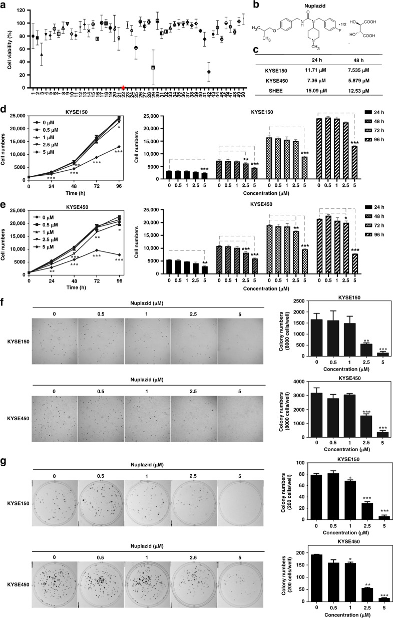
Fig. 1. Nuplaid inhibits the proliferation of ESCC cells.
a Nuplazid was screened from the drug library by cell cytotoxicity. b Chemical structure of Nuplazid. c IC50s of Nuplazid in KYSE150 cells, KYSE450 cells and SHEE cells. IC50s were calculated based on day 5 data of various doses of drug treatment. The KYSE150 cells (d) and KYSE450 cells (e) were treated with different doses of Nuplazid (0, 0.5, 1, 2.5 and 5 µM) and cell numbers were calculated at 0, 24, 48, 72 and 96 h by analysis at IN Cell Analyzer 6000. f KYSE150 cells and KYSE450 cells (8 × 10³/well) were exposed to different concentrations of Nuplazid (0, 0.5, 1, 2.5 and 5 µM) for 8 days. Colonies were counted for analysis by IN Cell Analyzer 6000 soft-agar program. g KYSE150 and KYSE450 cells (200/well) were treated with different concentrations of Nuplazid (0, 0.5, 1, 2.5 and 5 µM) and incubated for 8 days. Colonies were detected using the crystal violet stain assay. All data are shown as means ± S.D. The asterisks (*, **, ***) indicate a significant decrease (p < 0.05, p < 0.01, p < 0.001, respectively).

