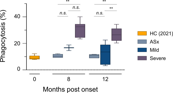
Figure 2.
In vitro phagocytic capability assays according to the severity of illness. ADCP assays with THP-1 cells as effectors and RBD-coated fluorescent, carboxylate-modified 1 μm red (580/605 nm, F8821) bead as targets (effector: target ratio of 10:1). RBD-coated red beads were pre-incubated with the diluted plasma for 20 min and then incubated with THP-1 cells for 2 h. Phagocytic activities were determined by flow cytometric analysis. % of Phagocytosis = numbers of red (580/605 nm) − positive THP-1 cells/numbers of total THP-1 cells × 100. Results are representative data from three independent experiments. Statistical analysis was performed using the Kruskal–Wallis rank-sum test with Dunn’s post hoc test in GraphPad Prism (n.s.: P > 0.05, **P < 0.01).

