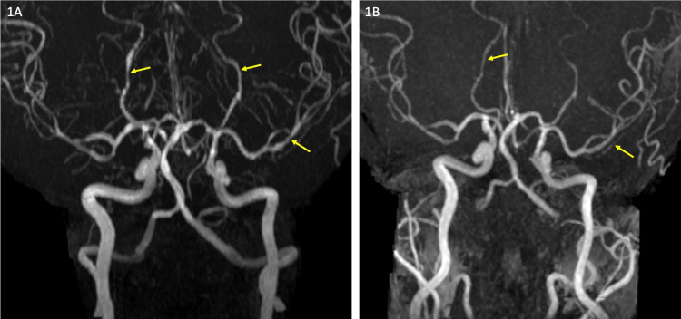
Figure 1. Magnetic resonance angiogram.
(A) Maximum intensity reconstruction from time of flight magnetic resonance angiogram (MRA) showing segmental irregularity and narrowing involving the distal bilateral posterior cerebral arteries and left middle cerebral artery (arrows). (B) Follow-up MRA after six weeks shows complete resolution of the vascular abnormality.

