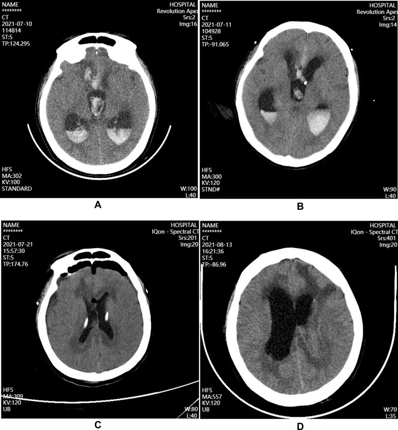
Figure 1.
Brain CT after admission in patient with intracranial A.baumannii infection. (A) Before the treatment on July 10, large high-density shadows can be seen on the right frontal lobe and lateral ventricle; (B) After the ventricular drainage on July 11, the right frontal lobe hemorrhage is slightly smaller than before; (C) During the treatment on July 21, ventricular hemorrhage and hydrocephalus was better than before; (D) During the treatment on August 13, bilateral lateral ventricle was dilated, and hydrocephalus progressed more than before.

